Empower confidence through evidence-based accuracy
Depend on the anatomy software solution trusted by millions worldwide
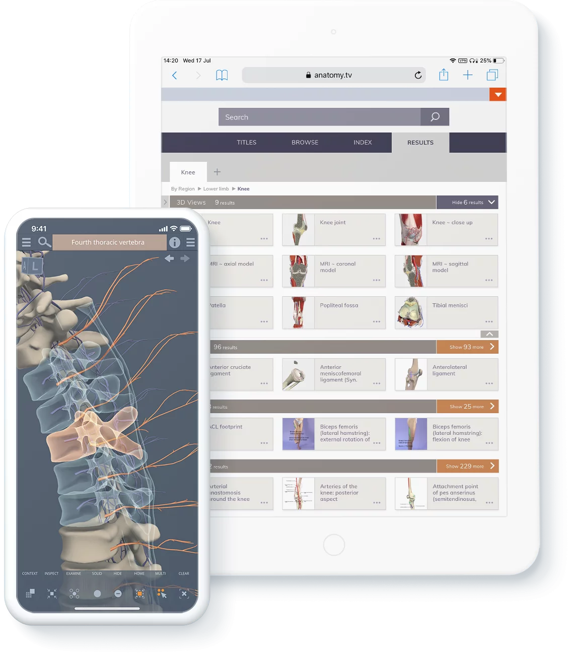
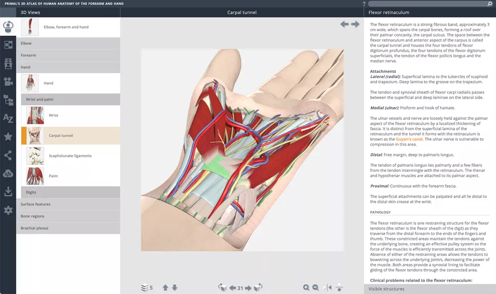
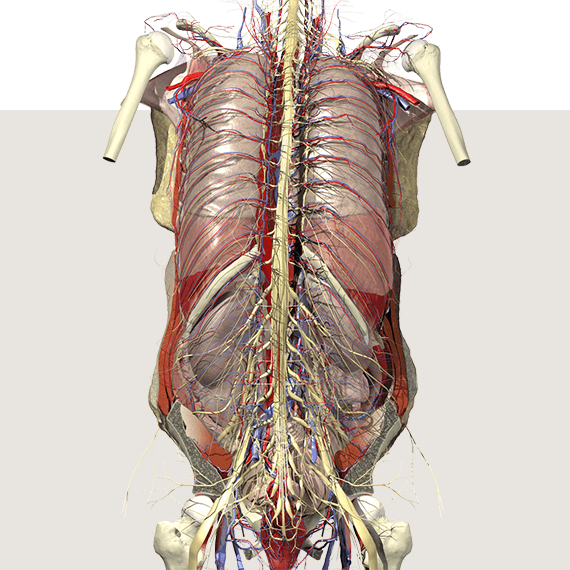
Primal Pictures and the Anatomy.tv platform – along with its mobile apps – is the world’s most detailed, accurate and evidence-based 3D reconstruction of human anatomy. Primal’s experts produced our digital model using real scan and imaging data. Advanced academic research and hundreds of thousands of development hours underpin its creation, which is exhaustively peer reviewed so you can teach and learn with confidence.